المدة الزمنية 8:22
Anatomy of Uterus - Parts and position, Blood supply, Nerve supply, Lymphatic drainage - Part 1
تم نشره في 2023/05/15
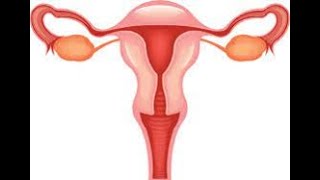
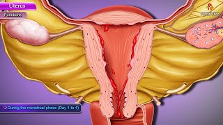
𝐒𝐮𝐛𝐬𝐜𝐫𝐢𝐛𝐞 𝗙𝐨𝐫 𝗠𝐨𝐫𝐞 𝗜𝐧𝐟𝐨𝐫𝐦𝐚𝐭𝐢𝐨𝐧 𝐨𝐧 𝗛𝐞𝐚𝐥𝐭𝐡 👩⚕ 𝐚𝐧𝐝 𝗠𝐞𝐝𝐢𝐜𝐢𝐧𝐞💉🩺💊 𝐘𝐨𝐮𝐭𝐮𝐛𝐞 : /@ DrAishwaryaKelkar 📌𝗙𝗮𝗰𝗲𝗯𝗼𝗼𝗸 : https://www.facebook.com/draishwaryakelkar 📌𝗧𝘄𝗶𝘁𝘁𝗲𝗿: https://twitter.com/AishwayaDr 📌𝗜𝗻𝘀𝘁𝗮𝗴𝗿𝗮𝗺 : https://www.instagram.com/clinical.learning/ Anatomy of Uterus - Parts and position, Blood supply, Nerve supply, Lymphatic drainage - Part 1 Parts and Position: ------------------------------ The uterus consists of several anatomical parts: Fundus: The uppermost portion of the uterus, which is rounded and convex. Body: The main central portion of the uterus, located between the fundus and the cervix. Cervix: The lower, cylindrical part of the uterus that projects into the vagina. It can be divided into two parts: the endocervix (lining the cervical canal) and the ectocervix (extending into the vagina). Internal Os: The opening at the upper end of the cervix that communicates with the uterine cavity. External Os: The opening at the lower end of the cervix that connects the cervix with the vagina. Blood Supply: --------------------- The blood supply to the uterus is provided by two main sets of arteries: Uterine Arteries: These arteries arise from the internal iliac arteries and are the primary blood supply to the uterus. They branch into arcuate arteries within the myometrium, which further give rise to radial arteries. The radial arteries supply the myometrium and the endometrium. Ovarian Arteries: These arteries arise from the abdominal aorta and contribute to the blood supply of the uterus. They anastomose with the uterine arteries. Nerve Supply: --------------------- The uterus receives its nerve supply from both the sympathetic and parasympathetic divisions of the autonomic nervous system. The nerves that innervate the uterus are derived from the uterovaginal plexus. Sympathetic fibers originate from the lumbar splanchnic nerves (T11-L2), while parasympathetic fibers arise from the pelvic splanchnic nerves (S2-S4). The autonomic nerve supply regulates the contractility of the uterus and controls blood vessel constriction or dilation. Lymphatic Drainage: -------------------------------- The lymphatic drainage of the uterus occurs via the following lymph nodes: Uterine Lymph Nodes: These lymph nodes are located along the uterine arteries and receive lymphatic drainage from the body of the uterus. External Iliac Lymph Nodes: Lymphatic vessels from the cervix and upper vagina drain into the external iliac lymph nodes. Sacral Lymph Nodes: Lymphatic vessels from the lower part of the vagina and the cervix drain into the sacral lymph nodes. Histology of the Uterus: ------------------------------------- The uterus is composed of three main layers: Endometrium: The innermost layer of the uterus that undergoes cyclic changes during the menstrual cycle. It consists of two layers: the functional layer (stratum functionalis), which sheds during menstruation, and the basal layer (stratum basalis), which regenerates the functional layer. Myometrium: The middle and thickest layer of the uterus composed of smooth muscle fibers. It is responsible for uterine contractions during labor and contributes to the expulsion of menstrual blood during menstruation. Perimetrium: The outermost layer of the uterus, which is a serous membrane that covers the uterus's surface. The histological composition of the uterus can vary depending on the stage of the menstrual cycle, pregnancy, or pathological conditions. #GrossAnatomy #Uterus #FemaleReproductiveSystem #AnatomyOfUterus #UterineStructure #UterinePosition #BloodSupply #NerveSupply #LymphaticDrainage #Histology #UterineLayers #UterusFunction #UterusExamination #UterineDisorders #UterinePathology #UterineSurgery #Physiology #Development #HormonalRegulation #MenstrualCycle #usmle #usmlestep1 #mbbs #uterusanatomy
الفئة
عرض المزيد


تعليقات - 3
مقاطع الفيديو ذات الصلة على Anatomy of Uterus - Parts and position, Blood supply, Nerve supply, Lymphatic drainage - Part 1:


![Moia File Virus Ransomware [.moia Removal and Decrypt] .moia Files](https://i.ytimg.com/vi/qoE7VSjFgvw/mqdefault.jpg)